Tuesday, March 25, 2014

Brain Tools

Tools we use to study the brain

Many tools are used to study the brain. Each works slightly differently.
For each of the following get an example of a brain image and a sentence or two about how it works or what it is used for.

CAT scan (computer axial tomography)
The CAT scan emits a series of narrow beams as it moves through. Many 2-D ray images are taken around a single axis of rotation and then placed together to create a 3-D image of thee brain.


Structural Magnetic Resonance Image
MRI scanning uses radio waves to produce images. The MRI scanner is a tube surrounded by a circular magnet.MRI scans are used as an accurate method of disease detection. It can view brain anatomy. 


Diffusion-Tensor MRI (DTI)
Diffusion MRI measures how water molecules diffuse through the body tissue. It is used too diagnose strokes, multiple sclerosis, etc. DTI allows researchers to map connectivity between brain areas. 


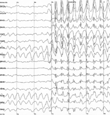
EEG (electroencephalograph)
 EEG uses electrodes to detect electrical activity in your brain since brain cells communicate via electrical impulses. EEG is used to diagnose epilepsy and other brain disorders. 


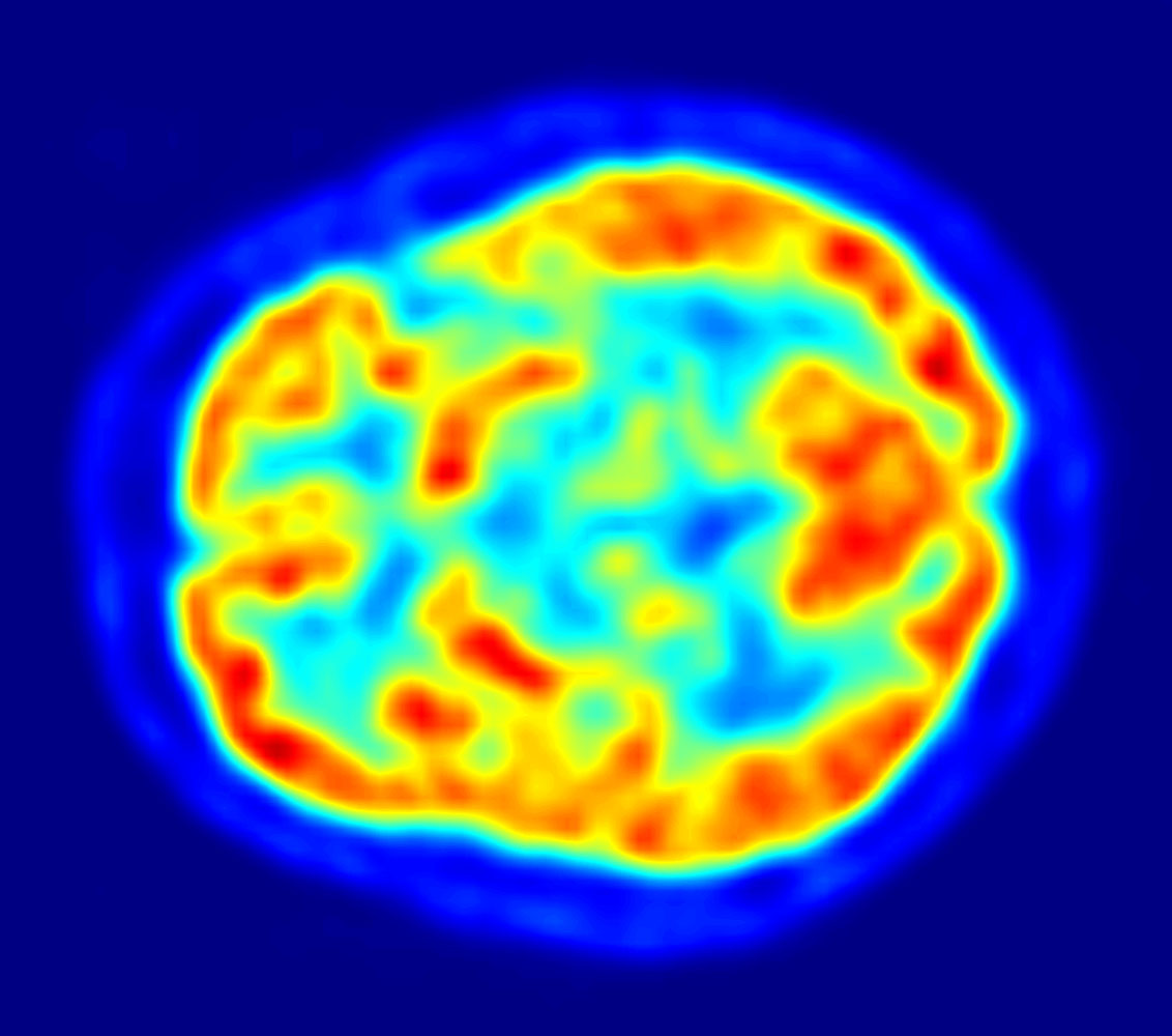
PET scan (Positron emission tomography)
The patient must consume radioactive material in his/her body. The PET scan tracks the radioactive material. More radioactive material often corresponds to areas of disease, etc. It is used in evaluating neurological problems.  


fMRI (functional MRI)
fMRI measures brain activity by detecting the changes in blood oxygenation and flow that occur in response to neural activity. fMRI shows parts of the brain that are involved in a particular mental process.


phMRI (pharamacological MRI)
phMRI uses drug stimuli to map brain function. It can provide information on neuro-receptor signaling and function. Drugs can elicit hemodynamic changes. 


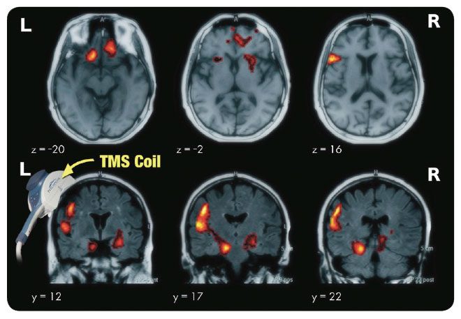
TMS (transcranial magnetic stimulation)
TMS uses magnetic fields to stimulate nerve cells in the brain to improve depression symptoms. A large electromagnetic coil is place on the head and sends electric currents. 
references: http://www.pbs.org/wnet/brain/scanning/
http://www.amenclinics.com/
http://faculty.washington.edu/chudler/image.html

Muscle Song

(to the tune of Twinkle Twinkle Little Star)

Muscles, Muscles in our body
so vital to our function
everywhere in our body
what would we do without you
Muscles, Muscles in our body

So many different types
cardiac smooth and skeletal
all in different areas
serving different functions
Muscles, Muscles in our body

Cardiac muscles in the heart
under involuntary control.
they appear to be striated
in the walls of the heart.
helping the heart beat everyday

Smooth muscles, in visceral organs
under involuntary control.
They appear spindle shaped,
contracting and relaxing,
working all over in the body.

Skeletal muscles, attached to bones
under voluntary control.
They appear to be striated,
stretching across bones and joints.
working with bones to give you movement

All these muscles are so useful
but how do they function?
myosin moving along filament
results in muscle contraction and release.
And that's the sliding filament theory.

Muscles, Muscles in our body
so vital to our function
everywhere in our body
what would we do without you
Muscles, Muscles in our body


Heart Sounds

Aortic Sclerosis
Aortic sclerosis heart beat is loud. It sounds like a slow horse galloping like bumBUm..bumBUM..bumBUM. It seems slow and steady.  Aortic sclerosis is when the trileaflet aortic valve calcifies and thickens. There is an absence of obstruction of ventricular outflow. As adults age, they tend to exhibit aortic sclerosis. Other factors that increase the risk of aortic sclerosis include being male, smoking, hypertension, high lipoprotein, and diabetes mellitus. LDL cholesterol lowering can slow the progression of aortic sclerosis. Aortic sclerosis can be an indicator of a coexisting coronary disease. Having aortic sclerosis may increase the risk of death from cardiovascular causes by 50%.

Aortic Stenosis (severe)
Aortic stenosis heart beart sounds like repetitive swift swinging of a baseball bat. It sounds deep, swift, and quick. It sounds like WOOP.WOOP.WOOP. It is louder and higher pitched. Aortic Stenosis is when the aortic valve does not fully open. It decreases the blood flow from the heart. The aortic valve becomes more narrow, making the left ventricle increase pressure to pump blood out. The muscles in the ventricle walls become thicker and causes chest pain. In severe cases, blood may back up into the lungs or prevent enough blood from reaching the brain or rest of the body. A common cause is rheumatic fever that develops after strep throat or scarlet fever. Another common cause is calcium deposits that narrow the valve.

Normal Heart (normal and unsplit)
It sounds normal and steady. It sounds like metronome going BUM...bum..BUM...bum..BUM..bum.  It goes 60 beats per minute. The first heart sound is a bit more intense than the second one. The first heart sound is produced by the closing of the mitral and tricuspid valve leaflets. The second heart sound is produced by the closing of the aortic and pulmonic valve leaflets.

Acute Pericarditis
It sounds like ocean waves crashing into another. It goes like WOOSH. WOOSH. WOOSH. The murmurs are caused by turbulent blood flow across incompetent waves, but a pericardial rub is caused by the two surfaces of the pericardial sack rubbing together. Pericarditis is a swelling and irritation of the pericardium, the thin membrane that surrounds your heart. Pericarditis causes chest pain. It is usually sudden and acute.

Mitral regurgitation (severe)
It sounds like an orca. It sounds like hoocrr..hoocrr..hoocrr. Severe mitral regurgitation is caused by the degeneration of the mitral valve leaflets. The first sound is normal but the second heart sound is widely split. It is characterized as a loud, rectangular, pansytolic murmur. Mitral regurgitation is the most common type of heart valve disorder. The mitral valve doesn’t close all the way and the blood flows backward into the upper heart chamber. There is a decrease in blood flow to the rest of the body and the heart tries harder to pump more blood. This may lead to congestive heart failure.

Atrial Septal Defect
Atrial septal defect sounds like someone knocking on the door. It goes like duh.DUH.duh.DUH. When a baby is in the womb, there’s an opening between the upper chambers and of the heart. It usually closes when the baby is born but if it doesn’t close the hole is considered an atrial septal defect. The hole allows blood to flow between the two heart chambers and pressure in the lungs may build up. There will be less oxygen in the blood that goes to the body.

Mediastinal Crunch
It sounds like the sound of the wind when you are recording a video on your phone. It sounds like a crunch. It is also known as Hamman’s sign. There is a crunching, rasping sound that is synchronous with the heartbeat. It is produced by the heart beating against air-filled tissues. IIt is caused by pneumomediastinum and is associated with tracheobronchial injury due to trauma.

Ebstein’s Anomaly
It sounds like someone snoring. Ebstein’s anomaly is a rare heart defect that is present at birth. The tricuspid valve, the valve between the right side chambers, doesn’t work properly. Blood leaks back through the valve, causing the heart to work less efficiently. It can lead to heart enlargement or heart failure.

Third Heart Sound
The third heart sound is an extra sound that creates a gallop cadence and sounds like “tennessee”. The third heart sound is heard early in diastole. It is very low frequency. To increase the intensity of the third heart sound, the patient should lie to his/her left side. It is heard on occasion in normal people with no cardiac pathology.

Exercise Heart Rate
Because exercise increases heart action, it increases the heart rate and the intensity of the first heart sound. This sounds like the first heart sound but louder and faster. The heart sounds are best heard using the diaphragm of the stethoscope.
References: http://www.practicalclinicalskills.com/heart-sounds-murmurs.aspx